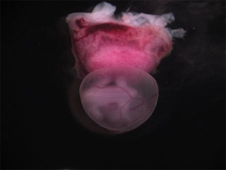
Our laboratory studies the uterus to advance gynecological and reproductive health and to inform mechanisms of regeneration. As one of the most dynamic and regenerative tissues in the adult body, the inner lining of the uterus or endometrium undergoes ~400 cycles of breakdown, growth, and scarless repair over the course of a woman’s lifetime. When perturbed, both reproductive and non-reproductive disorders can arise, including infertility, endometrial cancer, and endometriosis. Yet in spite of its central role in many human disorders and in reproduction, little is understood about the endometrium or its associated pathologies.


A central role of the uterus is reproduction. Pregnancy begins in eutherian mammals with implantation of an embryo into a receptive uterine endometrium. This maternal-fetal interface continues to form upon development of the embryonically-derived placenta, the organ connecting mother and fetus. Several conditions compromise the uterus’ ability to support a healthy pregnancy, including injury from prior procedures such as C-sections, which can lead to disorders such as placenta previa and placenta accreta. In addition, placental defects due to complications such as preeclampsia or intrauterine infection can also threaten the health and viability of both mother and fetus. Altogether, deficiencies in any of the steps of the intricate process of forming and maintaining the maternal-fetal interface can lead to maternal and/or fetal mortality and morbidities.
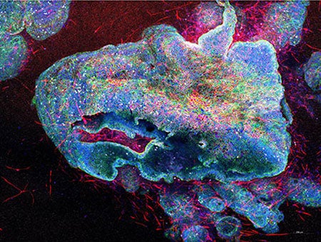
To address these challenges, our laboratory is using animal models and 3D organoids in combination with cutting-edge sequencing technologies and advanced microscopy to tackle the following major research directions:
- Uterine endometrial regeneration during the menstrual cycle
- Uterine wound healing following injury and its impact on subsequent pregnancies
- Development of novel 3D human organoid models of the uterine endometrium and placenta

